


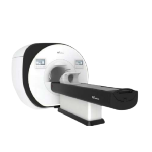
Description
Computed Tomography (CT) is a computerized imaging technique used in radiology. A rapidly rotating X-ray beam and detector are used to generate cross-sectional images – so-called slices – that form the volumetric and very detailed internal image of the body.
The external (visible) components of a CT scanner are the gantry, which includes a control panel, setup lasers and the bore along which the patient couch will traverse during the scan. While the idea of this imaging technique is more than 100 years old, it wasn’t until the early 1970s that sufficient computing power became reality. And although CT is now a very well-established and widely used diagnostic tool, there are still a lot of physics and engineering R&D activities being undertaken to improve and extend capabilities.
Currently the state-of-the-art detectors used in CT are energy integrating detectors (EID) but it looks like that use of a new upcoming technology based on photon counting detectors (PCD) might be the next leap ahead in CT imaging from a hardware perspective. While PCDs have become well-known and used for quite some time in SPECT (single photon emission computed tomography) systems, it was not possible until recently to also deploy them in CT Imaging systems.
EIDs use an indirect conversion for the signal creation. First the detector converts the X-ray energy into light, and then this light (i.e. multiple photons) is converted into an electrical current via photodiodes on the rear side of the detector. PCDs, on the other hand, allow for the detection – and energy discrimination – of single photons. The different detector material and the different method of data acquisition allows for energy-resolved CT images with the benefits of higher spatial resolution, decreased electronic noise and a potentially reduced radiation dose.
Reviews
There are no reviews yet.